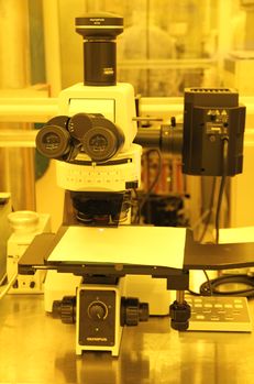
Fluorescence Microscope (Olympus MX51): Difference between revisions
Jump to navigation
Jump to search
Content deleted Content added
No edit summary |
|||
| (4 intermediate revisions by 2 users not shown) | |||
| Line 2: | Line 2: | ||
|picture=Fluorescence.jpg |
|picture=Fluorescence.jpg |
||
|type = Inspection, Test and Characterization |
|type = Inspection, Test and Characterization |
||
|super= |
|super= Demis D. John |
||
|location=Bay 6 |
|location=Bay 6 |
||
|description = |
|description = Optical Microscope |
||
|manufacturer = Olympus |
|manufacturer = Olympus |
||
|model = MX51 |
|||
|materials = |
|materials = |
||
|toolid= |
|toolid= |
||
}} |
}} |
||
=About= |
=About= |
||
System for general purpose imaging. The system has bright field, dark-field, |
System for general purpose imaging. The system has bright field, dark-field, DIC imaging mode and Three fluorescence filters. Screen captures and calibrated measurements can be made through use of native Olympus' Streams Essential Software, installed on the attached computer. |
||
=Detailed Specifications= |
=Detailed Specifications= |
||
* Native ''Olympus Streams Essentials'' software & Camera: |
|||
** Digital image capture & calibrated measurement (calibrations locked) |
|||
* Objectives: 5x, 10x, 20x, 50x, 100x, 150x |
|||
* Reflected (Episcopic) & Transmitted (Diascopic) Illumination modes |
|||
* Filters via rotatable selector: |
|||
** '''BF''': Bright Field normal top-illuminated imaging |
|||
** '''DF''': Dark-Field imaging |
|||
** '''DIC''': Differential Interference Contrast "Nomarski" Imaging. Also requires "DIC-R" prism to be inserted. Polarization manually selected on DIC-R prism. |
|||
* Fluorescence Filters (see [https://www.olympus-lifescience.com/en/microscope-resource/primer/techniques/fluorescence/anatomy/fluoromicroanatomy/ Olympus tutorial]): |
|||
** '''''You must contact Tony Bosch for training on Fluorescence before using this feature!!!''''' |
|||
** '''WB''': "Blue" Fluorescence Filter. Excitation: 450-480nm / Dichroic 500nm / Barrier 515nm+ |
|||
** '''WG''': "Green" Fluorescence Filter. Excitation: 510-550nm / Dichroic 570nm / Barrier 590nm+ |
|||
** '''WU''': "Ultraviolet" Fluorescence Filter. Excitation: 330-385nm / Dichroic 400nm / Barrier 420nm+ |
|||
=Documentation= |
=Documentation= |
||
Latest revision as of 18:09, 15 April 2020
|
About
System for general purpose imaging. The system has bright field, dark-field, DIC imaging mode and Three fluorescence filters. Screen captures and calibrated measurements can be made through use of native Olympus' Streams Essential Software, installed on the attached computer.
Detailed Specifications
- Native Olympus Streams Essentials software & Camera:
- Digital image capture & calibrated measurement (calibrations locked)
- Objectives: 5x, 10x, 20x, 50x, 100x, 150x
- Reflected (Episcopic) & Transmitted (Diascopic) Illumination modes
- Filters via rotatable selector:
- BF: Bright Field normal top-illuminated imaging
- DF: Dark-Field imaging
- DIC: Differential Interference Contrast "Nomarski" Imaging. Also requires "DIC-R" prism to be inserted. Polarization manually selected on DIC-R prism.
- Fluorescence Filters (see Olympus tutorial):
- You must contact Tony Bosch for training on Fluorescence before using this feature!!!
- WB: "Blue" Fluorescence Filter. Excitation: 450-480nm / Dichroic 500nm / Barrier 515nm+
- WG: "Green" Fluorescence Filter. Excitation: 510-550nm / Dichroic 570nm / Barrier 590nm+
- WU: "Ultraviolet" Fluorescence Filter. Excitation: 330-385nm / Dichroic 400nm / Barrier 420nm+
