Microscopes
|
Procedures & Tools
Microscope Training Guide
Use the guide linked above for general usage info & the many advanced features available on most of our microscopes. New users are encouraged to read this, and also request in-person training by Demis.
Image Analysis Software
Many of our microscopes have cameras and software for image capture. If you know the microscope and objective used for acquiring a photo, you can make calibrated measurements on the photos at your own desktop using the following software:
- AmScope Software - microscope image analysis software
- AmScope Calibration File containing calibrations for all NanoFab microscopes: Download Here
- Also available on Nanofiles-SFTP / Manuals / Amscope
- FIJI - scientific image anaylsis software
- The Microscope Measurement Tools plugin has pre-configured calibrations for NanoFab microscopes & SEMs, and allows you to draw length measurements.
- Calibrations in this plugin repository are out of date as of microscope upgrades in 2019.
- There are many other useful plugins, for particle counting, creating animations etc.
- The Microscope Measurement Tools plugin has pre-configured calibrations for NanoFab microscopes & SEMs, and allows you to draw length measurements.
Nikon
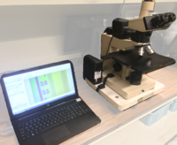
The Nikon scopes have 3rd party cameras that are connected to computers via AM Scope software which allows users to digitally capture images through the microscope and make calibrated measurements.
Microscope #2: Nikon Optiphot 200 (Bay 7)
- Trinocular: Ocular Binoc. + Camera (Simultaneous)
- Objectives: 5x, 10x, 20x, 50x, 100x, 150x
- Filters: Green, ND
- Bright/Dark Field
- Differential Interference Contrast (DIC/Nomarski)
- DIC only available on 100x and 150x mags due to image quality reduction at low mags due to DIC prisms which were removed for low mags.
- Top Reflected Illumination (Episcopic)
- AMScope 14MP Cameras with calibrated software for measurements. Software available for desktop measurements. Link here.
Microscope #3: Nikon Eclipse L200 (Bay 6)
- Manual for Nikon Eclipse L200D
- Trinocular: Binoc. + Camera (Mutually Exclusive)
- Objectives: 5x, 10x, 20x, 50x, 100x, 150x
- Filters: Green, ND
- Bright/Dark Field
- Differential Interference Contrast (DIC/Nomarski)
- Top Reflected Illumination (Episcopic)
- AMScope 14MP Cameras with calibrated software for measurements. Software available for desktop measurements. Link here.
Microscope #4: Nikon Eclipse L200D (Bay 6)
- Trinocular: Binoc. + Camera (Mutually Exclusive)
- Objectives: 5x, 10x, 20x, 50x, 100x, 150x
- Filters: Green, ND
- Bright/Dark Field
- Differential Interference Contrast (DIC/Nomarski)
- Top Reflected (Episcopic) & Bottom Transmission (Diascopic) Illumination
- AMScope 14MP Cameras with calibrated software for measurements. Software available for desktop measurements. Link here.
Olympus
The lab has two general purpose Olympus microscopes, and two specialized microscopes that require training to use, although both of these can also be used for general imaging.

Microscope #1: Olympus BHMJL (Char. Lab, Room 1111)
- Trinocular: Ocular Binoc. + Camera (Exclusive)
- LED Illuminator, Variable
- Objectives: 10x, 20x, 50x, 100x
- Bright/Dark Field
- Top Reflected (Episcopic) & Bottom Transmission (Diascopic) Illumination
- AMScope 14MP Cameras with calibrated software for measurements. Software available for offline measurements: Link here.
- User Manual: Olympus BHM Microscopes - Instruction Manual (PDF)
Microscope #5: Fluorescence Microscope: Olympus MX51 (Bay 6)
- See the wiki page for the Olympus MX51 for full details
- Trinocular: Binoc. + Camera (Simultaneous)
- Native Olympus Stream Software:
- Photo/video capture
- Calibrated measurement (calibrations locked)
- Objectives: 5x, 10x, 20x, 50x, 100x, 150x
- Filters: Green, ND
- Bright/Dark Field
- Differential Interference Contrast (DIC/Nomarski)
- Top (Episcopic) & Bottom (Diascopic) Illumination
- Three Fluorescence Filters (requires training, see main tool page for specs.)
Microscope #6: DUV Microscope: Olympus MX61A-DUV (Bay 7)
Please see the main tool page for detailed info on this microscope: Deep UV Optical Microscope (Olympus)
YOU ARE REQUIRED TO GET TRAINED on this tool before you are allowed to use it! Please contact the Tool Owner to get training.
- Motorized Stage + Objective Turret
- Trinocular: Binoc. + Camera (Simultaneous)
- Objectives: 5x, 10x, 20, 50x, 100x, DUV-100x
- Filters: to be added
- Native Olympus MX61 Software control & Camera
- Calibrated measurements (calibrations locked)
- Z (focus) measurement via motorized stage height
- Deep-UV Light source + DUV Camera
- DUV-100x sub-micron imaging/measurement
Olympus LEXT Confocal Microscope (Bay 4)
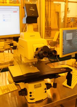
See the main tool page for complete info: Laser Scanning Confocal M-scope (Olympus LEXT)
YOU ARE REQUIRED TO GET TRAINED on this tool before you are allowed to use it! Please contact the Tool Owner to get training.
- Motorized stage + Objective Turret
- 100mm wafer stage
- Native Olympus OLS2000 Software & Built-In Camera:
- Calibrated measurement (calibrations locked)
- Image stitching capabilities
- 3D Laser-Scanning Confocal Microscopy capability:
- 3D Topographical measurement (optical profilometry)
- Surface roughness estimations (large roughness)
- Thin-Film Film-Thickness Measurements (thicker films)
Procedures & Documentation
- Microscope Training - General procedures and info for using our microscopes
